
mammography
Our Services
mammography
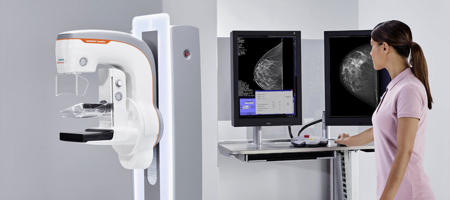
Mammography
Mammography is the taking of detailed images of the breast on film with the help of a special low-dose x-ray (X-Ray). In mammography, low-dose X-rays, high-contrast and high-density films and specially designed x-ray devices are used. Early diagnosis is very important for the success of breast cancer treatment. Mammography plays the most important role in early breast cancer diagnosis. The United States Food and Drug Administration estimates that 85% to 90% of lumps in women over the age of 50 can be detected by mammography 2 years before they reach a noticeable size. The benefits of mammography far outweigh its potential risks and discomfort.
Mammography can display changes in the breast even when they cannot be felt by the woman or her doctor. Once the mass is found, mammography can be used to determine whether it is cancer. For this purpose, a biopsy can be performed with the help of mammography. Biopsy is the process of removing suspicious tissue and determining whether it is cancerous under laboratory conditions. If an abnormality is found, a biopsy performed with a special type of mammography (Stereotactic Mammography) or with the help of ultrasound can be used to confirm the diagnosis.
Types of Mammography
Mammography can be divided into two: for control and diagnostic purposes;
• Control mammography is used to diagnose possible breast cancers at an early stage in women who have no complaints. When control mammography is performed regularly, it increases the possibility of early diagnosis and significantly increases the chance of success of the treatment. It is recommended that every woman over the age of 40 have a check-up mammogram once a year. Women who are considered to have a relatively higher risk can start this practice at an earlier age with the recommendation of their doctor.
• Diagnostic mammography is a method used for women with breast-related complaints such as the presence of a mass or breast discharge, and for women with abnormal formations in their control mammography. Diagnostic mammography is a more detailed procedure and therefore takes longer than control mammography. Diagnostic mammography is performed to determine the exact location and size of the suspicious tissue and to image the surrounding tissue and lymph nodes. Women who have previously had breast cancer and women who have breast prostheses (silicone, etc.) can be viewed from more angles when having a diagnostic mammography.
How is Mammography Done?
While mammography is being performed, the specialist positions the patient in the required position and views each breast separately. For each breast shot, the breast is carefully placed on the film carrier plates and gently pressed between the two plates. These sheets are usually made of clear fiberglass or other clear plastics. This compression causes the breast to flatten, making more successful visualization and examination of the tissue possible. The amount of radiation received by the tissue also decreases.
At some mammography centers, specialists may place adhesive locators on the breast skin before mammography. The purpose of this marking may be to identify formations that are not related to cancer, such as wounds, but may lead to incorrect evaluations, or it may be to mark potential areas such as a found mass that causes mammography. In some mammography centers, a marker is always placed on the nipple, and radiologists use this marker to locate their findings.
While mammography is being performed, an X-ray source is turned on and the resulting X-rays pass through the compressed breast and reach the film cassette under the breast. A special phosphor layer in the film cassette creates light photons in proportion to the X-rays falling on it, and these light photons are recorded by the x-ray film and a darkening occurs in the film. Since X-rays pass through tissues of different densities and types at different rates, the internal structure of the breast is imaged. This two-stage imaging method produces a very detailed image of the breast with the least possible amount of radiation, as it is performed using high-sensitivity x-rays and low-energy X-rays. Processed mammography films are evaluated by radiologists. In their evaluations, radiologists compare mammograms of both breasts with each other and new mammograms with old ones. In the evaluation, radiologists look at the shadows and distribution of breast tissue density.
Mammography is like fingerprints, it varies a lot from woman to woman, in fact, no mammogram is similar to another. For this reason, it is of great importance to take not only the reports of previous checks but also the mammography films taken during these checks. Small differences between previously taken mammography films and newly taken ones can enable cancer to be diagnosed at an early stage.
Breasts are composed of fat, connective tissue and glands. Breast masses, including benign and cancerous lesions, appear as white areas on the mammography film. Fat appears black on mammography film. Everything else (glands, connective tissue, tumors, other significant abnormalities such as microcalcification) appears as varying levels of white on the mammography film.
Compression of the breast during mammography
During mammography, the breast is compressed a little so that maximum tissue can be visualized. Although compression of the breast may cause some discomfort, this will only subside within the short period of time required for the mammogram. You may feel a hard squeeze and pressure during the mammogram, but this should not be painful. If you experience pain, you should notify the mammography technician. Reasons why the breast is compressed during mammography;
• When the breast is compressed, the breast tissue spreads over a larger area, overlaps are minimized, and therefore the anatomy of the breast and possible abnormalities can be visualized better. For example, inadequate compression leads to poor visualization of microcalcifications, which are tiny calcium deposits and are often an early sign of breast cancer.
• The possibility of shadows of normal structures overlapping and creating a suspicious image is reduced.
• Since a thinner tissue will be examined, less X-ray is required.
• By preventing the nozzle from moving, images are prevented from blurring.
• It prevents the X-ray from scattering within the thick tissue and reducing the image quality.
Images taken during mammography
During control mammography, the breasts are viewed separately. During this process;
• Typically from above (cranial-caudal view, CC)
• and, at an angle, obliquely (mediolateral-oblique, MLO)
image is taken.
In mammography performed for diagnostic purposes, the breasts are viewed separately. During this procedure, in addition to the images taken during control mammography, additional images related to the problem are taken. Among these;
• Lateromedial (LM) view taken from the outside to the inside
• Mediolateral (ML) image taken from the middle of the breast to the outside
• Magnified CC image taken from above
• There are images taken by compressing the problem area.
In addition, sometimes a "valley view" is taken to view the inside of both breasts. While this image is taken, both breasts are placed in the imaging location and the area between them and the inward-facing parts are imaged.
When abnormally dense tissues are detected in these images, more detailed imaging procedures are performed.
Mammography applications can be performed as “conventional” and “digital”. The startup procedures for both applications are the same. In other words, compression is applied to the breast (breasts are compressed) and an image is obtained using X-ray. However, image information is recorded on x-ray film in conventional mammography, and on the computer environment in digital mammography.
Conversion of analog images (gray scale information that forms a normal x-ray film) into digital information is done by 3 methods; use of film digitizer, computed radiography (CR) and digital radiography (DR).
Digitalization of plain graphs: It is the conversion of conventional plain graphs into digital data using a digitizer and is the least efficient method. It is reported that this method is useful in the transition from film-based system to PACS in large departments. However, since the loss of detail in mammograms will be high, it is not preferred in routine use.
CR : The CR technique is a technique that uses conventional radiography equipment to obtain digital data. Instead of conventional film, here a charged plate receives radiation. This plate then creates a digital image.
DR: It is a method in which electronic detectors (receivers) are used that directly convert the radiation passing through the patient into digital information. Although it is the most expensive among the three methods, it is the most practical way to obtain digital data in departments with high patient load.
The great progress made in today's computer technology has enabled digital radiological systems to develop at the same pace, and many different digital radiological methods have been developed. Digitalization of image data has provided great benefits in both archiving and image reprocessing, which is called reconstruction. In addition, the transfer of the obtained images from the place where the shooting was carried out to different centers through "network" communications enabled the evaluation and interpretation of the image data via computers.