
ultrasonography
Our Services
ultrasonography
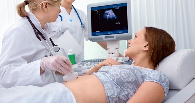
Ultrasonography is the imaging of organs and other structures within the body using high-frequency sound waves.
Hearing, one of our five senses, is provided by "sound". Humans cannot hear every sound; It can only hear certain sounds between 20-20000 Hertz. Sounds well above 20000 Hertz, which is the sound limit that humans cannot hear, are used in medicine. This method is called "Ultra Sono Graph" (USG). Since the sound we know is used even though its frequencies are very high, The examination does not cause any harm to the body. Harmful radiation such as X-rays etc. is not used.
An ultrasound device has two main parts: the main unit and the probe. The ultrasound piece placed on the body area examined during the examination is called "probe". High frequency sounds are converted into electrical energy through the transducer inside the probe. In the same way, the energy created by the sound waves coming from the body is converted into electrical energy through the transducer. These changes take place in the piazoelectric crystals inside the probe. Thus, the sound waves coming from the body are converted into images and made visible on the TV screen. The image created is called "sonogram". These images can be saved instantly with a printer.
Ultrasonography is most useful in examining organs and structures that contain fluid. Solid structures such as bones and air-filled organs such as the lungs do not appear well on ultrasound (since they do not contain much fluid). However, masses and solid formations in organs that contain fluid can be observed. Ultrasonography is used in various areas: in examining the abdominal organs (abdominal USG), in gynecological diseases (gynecological USG), in pregnancy and birth follow-up (obstetric USG), in examining heart functions and structure (echocardiography), in examining breast tissue (mammography), in examining the vessels. -In the examination of organs and structures such as thyroid-testis-eye etc... Different ultrasound devices and probes are used according to the location and characteristics of the organ examined. In ultrasonography, the probe is generally moved over the body (as in abdominal USG). It is performed by inserting the probe into the body. There are also types of USG: Transvaginal, transrectal etc.
PREPARATIONS TO BE MADE BEFORE THE EXAMINATION:
Except for emergencies, the patient must be hungry 12 hours in advance, take medication the night before, etc. (for upper abdominal USG), a fat-free diet should be followed one day before the test, (for kidney, bladder and gynecological USG) the bladder should be completely filled by drinking 1.5-2.5 liters of liquid (water) before the test. The examination is usually performed in the morning. If these conditions are met, it can be done at any time of the day.
APPLICATION (PERFORMANCE OF THE EXAMINATION):
An ultrasound image of the abdomen or any part of the body is obtained by taking various sections of that area. The patient lies first on his back, then (if necessary) on his stomach and on his side; The ultrasound probe is contacted to the body at different angles. Gel is applied to the body surface to prevent ultrasound waves from being lost between the body and the probe. The gel forms a bridge between the probe and the body.
ORGANS EXAMINED:
Examining the abdominal organs with ultrasound is called "abdominal ultrasonography". In medicine, the word abdomen is usually used instead of abdomen. The Latin word is abdomen.
Abdominal Ultrasonography is divided into two depending on the abdominal area where it is performed:
- Upper Abdominal Ultrasonography,
- Lower Abdominal Ultrasonography.
If both are performed together, it is called "Whole Abdominal Ultrasonography".
The following organs and structures are examined in upper abdominal ultrasonography:
Liver, gallbladder and bile ducts, kidneys and adrenal glands, spleen, pancreas, main vessels such as abdominal aorta and VCI, lower end of the esophagus.
In lower abdominal ultrasonography:
Bladder in men and women; prostate in men; In the woman, the uterus (womb), ovaries, tubes, and Douglas cavity are examined.
Abnormalities such as lymphadenopathy, mass (tumor, etc.), cyst, and fluid collection can also be detected in whole abdominal ultrasonography.
SITUATIONS WHICH REQUIRE ULTRASOUND:
Situations where "Whole Abdominal Ultrasonography" examination is required are as follows:
*Abdominal pain (may start a few hours ago or be persistent)
*Painful and/or bloody urination (may be accompanied by nausea and vomiting), intermittent urination
*Yellowing of the eyes and body, pain in the upper right side of the abdomen
*Having a family history of kidney disease, kidney stones, diabetes, etc.
*Continuous digestive complaints such as indigestion and nausea
*Continuous swelling of the abdomen
*In case of abdominal trauma such as a traffic accident or a hard blow to the abdomen (if there is a risk of internal organ rupture or bleeding)
*In patients with suspected abdominal tumor (mass), cyst or aortic enlargement
*If congenital abnormality or absence of intra-abdominal organs is considered
*To determine the size and structure of organs
*As part of general health screening (for check-up purposes)
DISEASES FOR WHICH ULTRASOUND IS MOST COMMON:
Ultrasound mostly investigates kidney stones, urinary tract infections, stones and infections in the gallbladder and bile ducts, spleen and liver enlargement, prostate enlargement (in men over 50 years of age), and gynecological diseases. Ultrasound is also frequently used in pregnancy follow-up.
FACTORS AFFECTING THE AUDIT:
- Contrast materials such as gas in the intestines, stool or barium remaining in the intestines from an examination performed in the last two days
- Patient movement during the examination (especially children)
- Excessive fatty tissue in obese people
- Presence of an open wound in the body area where ultrasound will be performed
ADVANCED EXAMINATIONS:
Further tests that may be required after ultrasonography are as follows:
- Blood tests
- Biopsy
- İVP, ERCP, PTK etc.
- Computed Tomography
- Magnetic resonance